Tag Archives: Small Times Magazine

April 19, 2011 — New MEMS, a 4-yr old segment of the MEMS market specifically for consumer electronics (CE) and mobile handsets, will grow by 157.4% in 2011, powering the expansion of the overall MEMS industry, according to new IHS iSuppli research.

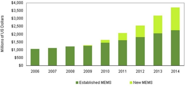
  2006 2007 2008 2009 2010 2011 2012 2013 2014
Established MEMS $1,049 $1,120 $1,214 $1,261 $1,460 $1,611 $1,808 $2,059 $2,245
New MEMS $0 $2 $6 $26 $178 $457 $740 $1,120 $1,465
Click to Enlarge

Figure. Revenue forecast for established MEMS vs. new MEMS in consumer and mobile (Millions of US Dollars). Source: IHS iSuppli Research, April 2011.

Revenue this year for new CE and mobile MEMS devices will reach $457.3 million, up by more than a factor of 2.5 from $177.6 million in 2010. A category including devices such as 3-axis gyroscopes and pico projectors, the new MEMS segment did not even exist in 2006. However, growth has been nothing short of explosive after the category was devised in 2007 to differentiate the segment from that of established MEMS. By 2014, new MEMS will generate revenue of $1.4 billion, as shown in the figure.

New MEMS enable motion-controlled video games, tablet navigation systems, pico projectors embedded in smart phones, and other exciting consumer products, said Jérémie Bouchaud, director and principal analyst MEMS and sensors for IHS. "This is driving fast revenue growth both for the new MEMS themselves and for the overall MEMS market."

Compared to the triple-digit growth rate of new CE and mobile MEMS, the established (accelerometers and single- or dual-axis gyroscopes) MEMS segment is poised to expand only 10.4% this year.

Established MEMS will continue to produce bigger revenue overall, but growth as a whole in the next three years will be confined to the range of 9 to 12%. Meanwhile, expansion rates for new CE and mobile MEMS will amount to a 31 to 62% from 2012 to 2014.

The net effect of such rapid growth is to increase the portion held by new MEMS of the total CE and mobile MEMS revenue pie. As a result, new MEMS in 2014 will account for 39.5% of overall revenue, a far cry from the 0.2 percent share in 2007 when the segment earned just $2.4 million. Without the revenue contribution of new MEMS, IHS believes, overall growth in the MEMS space will be dependable but underwhelming.

3-axis gyroscopes

Among new MEMS devices, 3-axis gyroscopes are most likely to take a star turn. Found in the likes of the iPhone 4 from Apple Inc. and the Move remote controller for the PlayStation 3 from Sony Corp., 3-axis gyroscopes feature prominently in applications for smart phones and gaming, with motion recognition functions benefiting greatly from the use of the new MEMS component.

3-axis gyroscopes also can be found in other consumer electronic devices, such as tablets like Apple’s iPad 2, Samsung Electronics’ Galaxy Tab as well as in every new tablet; the new 3DS handheld player from Nintendo Corp.; portable media players like Apple’s iPod Touch Fourth Generation (4G); and new smart phones from Samsung such as the Nexus or from LG Electronics like the Optimus.

Learn more in New MEMS Underpins Consumer Market Expansion: http://www.isuppli.com/MEMS-and-Sensors/Pages/New-MEMS-Underpins-Consumer-Market-Expansion.aspx?PRX

IHS iSuppli technology value chain research and advisory services range from electronic component research to device-specific application market forecasts, from teardown analysis to consumer electronics market trends and analysis and from display device and systems research to automotive telematics, navigation and safety systems research. More information is available at www.isuppli.com

April 19, 2011 — Plasmonics, a phenomenon in which the confinement of light in dimensions smaller than the wavelength of photons in free space make it possible to match the different length-scales associated with photonics and electronics in a single nanoscale device, has become one of the hottest fields in high-technology. However, to date plasmonic properties have been limited to nanostructures that feature interfaces between noble metals and dielectrics. Now, researchers with the U.S. Department of Energy (DOE)’s Lawrence Berkeley National Laboratory (Berkeley Lab) have shown that plasmonic properties can also be achieved in the semiconductor nanocrystals known as quantum dots.

"We have demonstrated well-defined localized surface plasmon resonances arising from p-type carriers in vacancy-doped semiconductor quantum dots that should allow for plasmonic sensing and manipulation of solid-state processes in single nanocrystals," says Berkeley Lab director Paul Alivisatos, a nanochemistry authority who led this research. "Our doped semiconductor quantum dots also open up the possibility of strongly coupling photonic and electronic properties, with implications for light harvesting, nonlinear optics, and quantum information processing."

Alivisatos is the corresponding author of a paper in the journal Nature Materials titled "Localized surface plasmon resonances arising from free carriers in doped quantum dots." Co-authoring the paper were Joseph Luther and Prashant Jain, along with Trevor Ewers.

Plasmonics could enable faster, higher-volume chip interconnects, microscope lenses that resolve nanoscale objects with visible light, highly efficient light-emitting diodes (LEDs), and supersensitive chemical and biological detectors. There is evidence that plasmonic materials can be used to bend light around an object, rendering that object invisible.

The plasmonic phenomenon was discovered in nanostructures at the interfaces between a noble metal, such as gold or silver, and a dielectric, such as air or glass. Directing an electromagnetic field at such an interface generates electronic surface waves that roll through the conduction electrons on a metal, like ripples spreading across the surface of a pond that has been plunked with a stone. Just as the energy in an electromagnetic field is carried in a quantized particle-like unit called a photon, the energy in such an electronic surface wave is carried in a quantized particle-like unit called a plasmon. The key to plasmonic properties is when the oscillation frequency between the plasmons and the incident photons matches, a phenomenon known as localized surface plasmon resonance (LSPR). Conventional scientific wisdom has held that LSPRs require a metal nanostructure, where the conduction electrons are not strongly attached to individual atoms or molecules. This has proved not to be the case.

Prashant Jain, a member of the Alivisatos research group and one of the lead authors of the Nature Materials paper, explains, "Our study represents a paradigm shift from metal nanoplasmonics as we’ve shown that, in principle, any nanostructure can exhibit LSPRs so long as the interface has an appreciable number of free charge carriers, either electrons or holes. By demonstrating LSPRs in doped quantum dots, we’ve extended the range of candidate materials for plasmonics to include semiconductors, and we’ve also merged the field of plasmonic nanostructures, which exhibit tunable photonic properties, with the field of quantum dots, which exhibit tunable electronic properties. Unlike a metal, the concentration of free charge carriers in a semiconductor can be actively controlled by doping, temperature, and/or phase transitions. Therefore, the frequency and intensity of LSPRs in dopable quantum dots can be dynamically tuned. The LSPRs of a metal, on the other hand, once engineered through a choice of nanostructure parameters, such as shape and size, is permanently locked-in."

Jain and his co-authors made their quantum dots from the semiconductor copper sulfide, a material that is known to support numerous copper-deficient stoichiometries. Initially, the copper sulfide nanocrystals were synthesized using a common hot injection method. While this yielded nanocrystals that were intrinsically self-doped with p-type charge carriers, there was no control over the amount of charge vacancies or carriers.

"We were able to overcome this limitation by using a room-temperature ion exchange method to synthesize the copper sulfide nanocrystals," Jain says. "This freezes the nanocrystals into a relatively vacancy-free state, which we can then dope in a controlled manner using common chemical oxidants."

By introducing enough free electrical charge carriers via dopants and vacancies, Jain and his colleagues were able to achieve LSPRs in the near-infrared range of the electromagnetic spectrum.

Jain envisions quantum dots as being integrated into a variety of future film- and chip-based photonic devices that can be actively switched or controlled, and also being applied to such optical applications as in vivo imaging.

In addition, the strong coupling that is possible between photonic and electronic modes in such doped quantum dots holds exciting potential for applications in solar photovoltaics and artificial photosynthesis. "In photovoltaic and artificial photosynthetic systems, light needs to be absorbed and channeled to generate energetic electrons and holes, which can then be used to make electricity or fuel," Jain says. "To be efficient, it is highly desirable that such systems exhibit an enhanced interaction of light with excitons. This is what a doped quantum dot with an LSPR mode could achieve."

The potential for strongly coupled electronic and photonic modes in doped quantum dots arises from the fact that semiconductor quantum dots allow for quantized electronic excitations (excitons), while LSPRs serve to strongly localize or confine light of specific frequencies within the quantum dot. The result is an enhanced exciton-light interaction. Since the LSPR frequency can be controlled by changing the doping level, and excitons can be tuned by quantum confinement, it should be possible to engineer doped quantum dots for harvesting the richest frequencies of light in the solar spectrum.

Quantum dot plasmonics also hold intriguing possibilities for future quantum communication and computation devices. "The use of single photons, in the form of quantized plasmons, would allow quantum systems to send information at nearly the speed of light, compared with the electron speed and resistance in classical systems," Jain says. "Doped quantum dots by providing strongly coupled quantized excitons and LSPRs and within the same nanostructure could serve as a source of single plasmons."

Jain and others in Alivsatos’ research group are now investigating the potential of doped quantum dots made from other semiconductors, such as copper selenide and germanium telluride, which also display tunable plasmonic or photonic resonances. Germanium telluride is of particular interest because it has phase change properties that are useful for memory storage devices.

"A long term goal is to generalize plasmonic phenomena to all doped quantum dots, whether heavily self-doped or extrinsically doped with relatively few impurities or vacancies," Jain says.

This research was supported by the DOE Office of Science.

Lawrence Berkeley National Laboratory addresses the world’s most urgent scientific challenges by advancing sustainable energy, protecting human health, creating new materials, and revealing the origin and fate of the universe. Founded in 1931, Berkeley Lab’s scientific expertise has been recognized with 12 Nobel prizes. The University of California manages Berkeley Lab for the U.S. Department of Energy’s Office of Science. For more, visit www.lbl.gov.

For more information about the research of Paul Alivisatos, visit the Website at http://www.cchem.berkeley.edu/pagrp/

For more information about the research of Prashant Jain, visit the Website at http://www.nanogold.org/

Follow Small Times on Twitter.com by clicking www.twitter.com/smalltimes. Or join our Facebook group
April 19, 2011 – BUSINESS WIRE — MicroVision (Nasdaq: MVIS), ultra-miniature display technology provider, opened a research and development center at Nanyang Technological University (NTU), Singapore. The research facility, located on NTU’s 200-hectare green campus, will focus on developing innovative breakthrough products using MicroVision’s PicoP Display technology.

MicroVision plans to staff the new R&D facility with up to 25 engineers by 2012 to work on advanced research and development projects, perform operational support functions, and build upon the company’s current portfolio of over 500 patents issued and pending. By collaborating with NTU, MicroVision aims to leverage the university’s strength and expertise in engineering, microelectronics, and materials science to conduct joint research and development with faculty and students. The alliance is also expected to facilitate the exchange of ideas between NTU staff and students and MicroVision personnel, as well as provide possible internship opportunities for NTU students.

MicroVision’s dedicated R&D center includes a customized laboratory at NTU’s Innovation Centre. The company will work directly with NTU’s School of Electrical and Electronic Engineering and the Division of Physics and Applied Physics.

The Memorandum of Understanding between NTU and MicroVision was signed by Professor Bertil Andersson, NTU’s President-Designate and Provost and Alexander Tokman, CEO and president, MicroVision.

Tokman called Singapore centrally located to MicroVision’s manufacturers and customers, and "a hub for exceptional technical talent and productivity," adding that the students at NTU proffer a wealth of new ideas and fresh thinking.

This is MicroVision’s first R&D center outside the United States. The company originally considered Taiwan, but decided on NTU citing expertise in engineering and computing and an excellent research infrastructure, said Andersson. MicroVision engineers will work side by side with NTU faculty and students to perform joint research into innovative imaging and display solutions.

MicroVision provides the PicoP display technology platform designed to enable next-generation display and imaging products for pico projectors, vehicle displays and wearable displays that interface with mobile devices. MicroVision has a history of collaborating with leading universities and research institutes across the globe, including Stanford and MIT (USA) and Fraunhofer Institute (Germany). For more information, visit the company’s website at www.microvision.com.

NTU has been rapidly ramping its research capabilities in the last few years and has established strong industry partners including Rolls-Royce, Robert Bosch, Thales, and Toray. A research-intensive public university, Nanyang Technological University (NTU) has 33,500 undergraduate and postgraduate students in the colleges of Engineering, Business, Science, and Humanities, Arts, & Social Sciences. In 2013, NTU will enroll the first batch of students at its new medical school, the Lee Kong Chian School of Medicine, which is set up jointly with Imperial College London. For more information, visit www.ntu.edu.sg

Follow Small Times on Twitter.com by clicking www.twitter.com/smalltimes. Or join our Facebook group

April 18, 2011 – BUSINESS WIRE — Agilent Technologies Inc. (NYSE:A) delivered a fully integrated 1.1-THz network analysis measurement solution to Japan’s Yamaguchi University. The solution will play a critical role in enabling the university to study metamaterials at THz frequencies.

Click to Enlarge

The THz region of the electromagnetic spectrum has enormous potential for high-data-rate communications, advanced electronic materials spectroscopy, space research, medicine, biology, surveillance and remote sensing. Despite its potential, the THz region remains one of the most unexplored areas of the spectrum. And, since high-power THz sources rely heavily on materials with advantageous properties in the THz frequency range, materials research is a critical component of modern THz systems.

Agilent’s integrated measurement solution addresses this challenge by enabling accurate measurement of THz signals in new materials. Such information is critical to the successful resolution of Yamaguchi University’s materials research.

Agilent’s solution features a fully calibrated, 750-GHz to 1.1-THz frequency extension module, the WR-01, from Virginia Diodes Inc., coupled with Agilent’s high-performance 50-GHz PNA-X vector network analyzer. Together, these tools allow for a fully calibrated, vector network analysis measurement with greater than 50dB of dynamic range.

Users can take advantage of the PNA-X’s calibration technology to make stable and repeatable THz measurements. The instrument’s dynamic range also ensures that users can easily achieve highly sensitive measurements. And, with the frequency extension module’s ability to operate across the 750-GHz to 1.-THz frequency range, users can now easily capture higher resolution images as well.

"At Virginia Diodes, our mission is to make the THz region of the electromagnetic spectrum as useful for scientific, military and commercial applications as the microwave and infrared bands are today," said Thomas Crowe, chief executive officer of Virginia Diodes. The WR-01 extender offers the dynamic range and bandwidth required for THz-calibrated vector network analyzer measurements, Crowe added.

"Working closely with Virginia Diodes, we were able to integrate the WR-01 extender into our PNA-X vector network analyzer, further extending its capabilities and enabling fast and accurate measurements at THz," said Gregg Peters, vice president and general manager of Agilent’s Component Test Division.

Agilent Technologies Inc. (NYSE: A) provides tools for chemical analysis, life sciences, electronics and communications. Information about Agilent is available at www.agilent.com. Agilent’s PNA-X vector network analyzer provides the industry’s widest range of measurement applications, from RF to millimeter wave. More information about the PNA-X is available at www.agilent.com/find/pna-x

Yamaguchi University is located in Ube-city, Japan. Its characterization of materials at THz frequencies is part of an ongoing effort within the school of science and engineering. For more information, go to http://www-ap.apsci.yamaguchi-u.ac.jp/groups/appliedphysicslaboratory/

Follow Small Times on Twitter.com by clicking www.twitter.com/smalltimes. Or join our Facebook group

April 15, 2011 — OPEN ENGINEERING, the Multiphysics branch of the SAMTECH Group, a leading European provider of simulation software and services, signed a cooperation agreement with the Russian University of Information Technologies, Mechanics and Optics (ITMO).

ITMO recognizes OPEN ENGINEERING, active in development of Multiphysics software (also known as numerical simulation programs) as a strategic partner to support its research program for innovative Russian industries and SMEs. Open Engineering software solutions will help ITMO’s customers to create their products on computers, to test them virtually and to improve and optimize them before producing the first prototype.

"This agreement rewards our policy of international expansion," explains Olivier Gramaccia, SAMTECH sales & marketing director.

SAMTECH provides computer aided engineering (CAE) software. SAMTECH develops and markets the general-purpose Finite Element Analysis code SAMCEF, the Multi-Disciplinary Optimization platform BOSS Quattro and the Open CAE Integration Framework CAESAM. Visit www.samtech.com.

OPEN ENGINEERING, part of the SAMTECH Group, supplies Multiphysics software for the CAE market. Their solutions are based on the Oofelie Multiphysics platform, optimized for large complex industrial 3D design work. Oofelie Multiphysics provides its users with unique capabilities to analyze industrial applications such as: sensors & actuators, MEMS & MOEMS and Fluid Structure Interaction problems. The company was set up in 2001 as a subsidiary of the SAMTECH Group and a spin-off company from the University of Liège (ULg). Visit http://www.open-engineering.com for more information.

The Saint Petersburg State University of Information Technologies, Mechanics and Optics (University ITMO) is one of the leading higher education institutions in Russia providing training in advanced science and technology. Visit http://en.ifmo.ru for details.

Follow Small Times on Twitter.com by clicking www.twitter.com/smalltimes. Or join our Facebook group
April 15, 2011 — University of Maryland researchers have discovered a way to control magnetic properties of graphene that could lead to powerful new applications in magnetic storage and magnetic random access memory.

The finding by a team of Maryland researchers, led by Physics Professor Michael S. Fuhrer of the UMD Center for Nanophysics and Advanced Materials is the latest of many amazing properties discovered for graphene.

Click to Enlarge

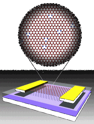
Image. Schematic of a graphene transistor showing graphene (red), gold electrodes (yellow), silicon dioxide (clear) and silicon substrate (black). Inset shows the graphene lattice with vacancy defects. Vacancies (missing atoms) are shown surrounded by blue carbon atoms. Graphic by Jianhao Chen and Michael S. Fuhrer, University of Maryland.

In their new graphene discovery, Fuhrer and his University of Maryland colleagues have found that missing atoms in graphene, called vacancies, act as tiny magnets — they have a "magnetic moment." These magnetic moments interact strongly with the electrons in graphene that carry electrical currents, giving rise to a significant extra electrical resistance at low temperature, known as the Kondo effect. The results appear in the paper "Tunable Kondo effect in graphene with defects" published this month in Nature Physics. Access the article here.

The Kondo effect is typically associated with adding tiny amounts of magnetic metal atoms, such as iron or nickel, to a non-magnetic metal, such as gold or copper. Finding the Kondo effect in graphene with vacancies was surprising for two reasons, according to Fuhrer.

"First, we were studying a system of nothing but carbon, without adding any traditionally magnetic impurities. Second, graphene has a very small electron density, which would be expected to make the Kondo effect appear only at extremely low temperatures," he said.

The team measured the characteristic temperature for the Kondo effect in graphene with vacancies to be as high as 90 Kelvin, which is comparable to that seen in metals with very high electron densities. Moreover the Kondo temperature can be tuned by the voltage on an electrical gate, an effect not seen in metals. They theorize that the same unusual properties of that result in graphene’s electrons acting as if they have no mass also make them interact very strongly with certain kinds of impurities, such as vacancies, leading to a strong Kondo effect at a relatively high temperature.

Fuhrer thinks that if vacancies in graphene could be arranged in just the right way, ferromagnetism could result. "Individual magnetic moments can be coupled together through the Kondo effect, forcing them all to line up in the same direction," he said. "The result would be a ferromagnet, like iron, but instead made only of carbon. Magnetism in graphene could lead to new types of nanoscale sensors of magnetic fields. And, when coupled with graphene’s tremendous electrical properties, magnetism in graphene could also have interesting applications in the area of spintronics, which uses the magnetic moment of the electron, instead of its electric charge, to represent the information in a computer.

"This opens the possibility of ‘defect engineering’ in graphene — plucking out atoms in the right places to design the magnetic properties you want," said Fuhrer.

Graphene conducts electricity at room temperature better than any other known material (a 2008 discovery by Fuhrer, et. al). Graphene is widely seen as having great, perhaps even revolutionary, potential for nanotechnology applications. The 2010 Nobel Prize in physics was awarded to scientists Konstantin Novoselov and Andre Geim for their 2004 discovery of how to make graphene.

This research was supported by grants from the National Science Foundation and the Office of Naval Research.

Research at the UMD Center for Nanophysics and Advanced Materials focuses on understanding the limits of graphene’s conductivity, what causes the scattering of its electrons, and how to make graphene more stable and reliable. This University of Maryland research is an interdisciplinary effort, involving investigative teams in nanotechnology, materials science and condensed matter physics. Michael Fuhrer leads the team that investigates the possibilities of graphene for electronic application, particularly exploring the potential of graphene’s high level of mobility and the promise that suggests for the material’s use in electrically conducting, transparent film. A team founded by renowned UMD physicist and materials scientist Ellen Williams leads research on surface science. Current experimentation focuses on determining the effects of the impurities in graphene, leading to an understanding of the material’s potential in a cleaner state. Maryland’s Physics Professor Sankar Das Sarma, Distinguished University Professor & Director of the Condensed Matter Theory Center, leads a team of post-doctoral researchers interested in understanding the theory behind the science of graphene and the research being done on its applications

April 15, 2011 — Electron microscopes use focused electron beams to make extremely small objects visible. By combining the microscope with a gas-injection system, material samples can be manipulated and nanometer-wide surface structures can be written. Empa researchers, together with scientists from EPFL, used this method to improve lasers.

The vertical cavity surface emitting laser (VCSEL), a semiconductor laser often used in data transmission for short-distance links like Gigabit Ethernet, exhibits one weakness: Because of the cylindrical structure in which the lasers are built up on the wafer, the polarization of the emitted light can sometimes change during operation. Stable polarization is necessary to reduce transmission errors and to use VCSELs in future silicon photonics.

Click to Enlarge

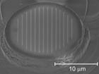
Image: The result is a nanostructure — for example, a polarization grating on a VCSEL (vertical cavity surface emitting laser). These are semiconductor lasers frequently used in optical data transmission.

"We’ve written flat grating structures on the VCSELs with an electron beam," says Ivo Utke, Empa researcher, in describing their solution, "and the gratings were effective in stabilizing the polarization."

Click to Enlarge

Image. The principle of the local deposition process, which is induced with a focused electron beam (FEBIP), is that molecules from a gas-injection system are deposited on the sample surface in a reversible manner. The focused electron beam dissociates adsorbed gas molecules. The resulting non-volatile compounds remain permanently on the sample.

Minimally invasive, direct FEBIP is suitable for prototyping nanocomponents to solve specific questions and problems in applied nanoelectronics, nanophotonics, and nanobiology. Suitable gas molecules are injected close to a sample which is already in the microscope’s vacuum chamber. These adsorb on the sample in a reversible manner.

The focused electron beam induces chemical reactions of the adsorbed gas molecules, but only at the spot where the beam strikes the surface. The resulting non-volatile molecular fragments then remain permanently on the sample while the volatile fragments are removed by the vacuum system. "With the help of a precisely positioned electron beam, it’s possible to remove or apply surface structures with nanometer precision and in virtually any desired three-dimensional shapes, explains Utke. "FEBIP could soon become a true nanofabrication platform for rapid prototyping of nanostructures in a minimally invasive way, without necessitating the large investment of a clean room."

The team was led by Utke, together with scientists from the Laboratory of Physics of Nanostructures at EPFL. The study has recently been published in the scientific journal Nanoscale as an advanced online publication. Access the article here.

Learn more at www.empa.ch

Follow Small Times on Twitter.com by clicking www.twitter.com/smalltimes. Or join our Facebook group
Gabriel Monette, Université de Montréal, Montreal, Canada and Marc Verhaegen, Photon etc., Montreal, Canada

Magneto-optical (MO) effects are used as tools for probing the magnetization reversal characteristics of a wide range of sample types, and as an effective way to modify the polarization of light via induced magnetization state in samples. In transparent samples, the Faraday effect (rotation of the polarization of light proportional to the magnetic induction field and optical path in the medium) can be used to elaborate Faraday rotators, a key element in the design of optical isolators [1]. Along with other MO effects such as Kerr measurements, they provide a non-destructive probe for in-situ measurements of samples, such as thin films.

Spectral dependence of the Faraday MO effect in the visible part of the electromagnetic spectrum along with temperature dependence measurements were performed on a semiconductor 2micron epilayer (GaP) grown with embedded metallic ferromagnetic nanoclusters (MnP). The confined geometry of the experiment — which necessitates a cryostat chamber with optical window placed within the pole gap of an electromagnet applying the DC magnetic field parallel to the normal of the sample — made it difficult to use a monochromatic light beam produced by filtering an incandescent light source with a standard monochromator.

Click to Enlarge

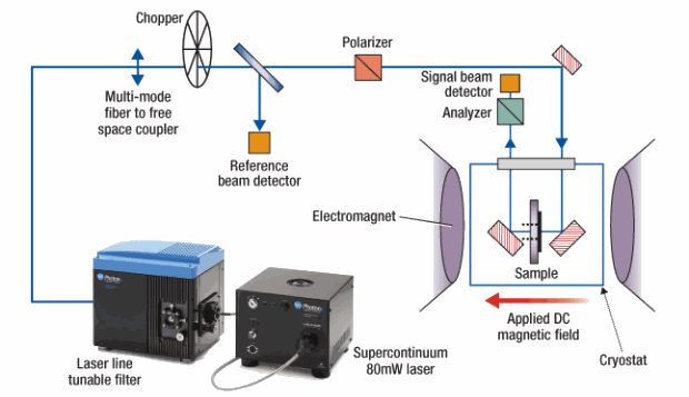
Figure 1. Experimental set up.

Instead, the team used a collimated tunable laser based on a Leukos (SP20) supercontinuum source and Photon etc’s Tunable Laser Line Filter. Figure 1 shows the experimental set up used to investigate the MO properties of the GaP:MnP in the Faraday configuration. The analyzer is mounted on a motorized rotation stage to allow tracking of the extinction condition as a function of temperature, wavelength and applied magnetic field. Hysteresis curves were obtained by rotating the analyzer at 45° with respect to the polarizer and sweeping the magnetic field from -400 to 400 mT. Small angles of rotation ensure linear variation in transmitted intensity as a function of the rotation angle of the polarization, or applied magnetic field. The source of the electromagnet, the temperature controller for the cryostat, the angular position of the analyzer, and the wavelength selection of the laser output (via Photon etc Tunable Laser Line Filter) are all computer controlled. The angle of rotation must be obtained independently for each applied field, wavelength, and temperature.

Click to Enlarge
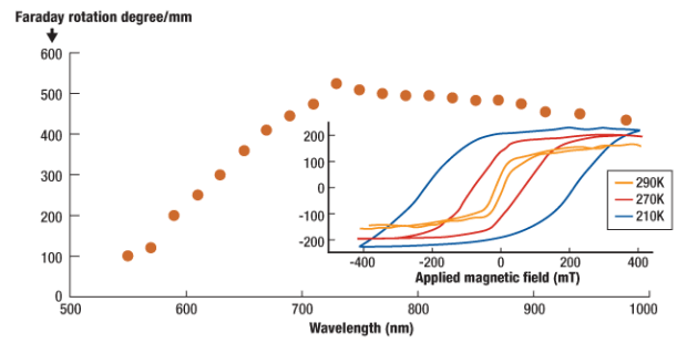
Figure 2. Faraday rotation vs wavelength.

The total Faraday rotation of the epilayer as a function of wavelength at a temperature of 220K is displayed in figure 2. The free carriers contribution of the GaP substrate has been carefully substracted. The GaP:MnP epilayer produces a maximum MO effect in the near infrared, whereas the substrate has a monotonic decrease in the MO effect as the wavelength is increased. The inset shows the Faraday rotation hysteresis curves at 210K, 270K and 290K at 655nm. The giant Faraday rotation in these systems has been reported [2]. The tunable laser allowed us to investigate the hysteresis signature of the MO Faraday effect at different wavelengths as well as different temperatures within the limited work space of the apparatus.

Future applications

Although this specific experiment and material (GaP:MnP) aim at a better understanding of the underlying physics of effective medium, i.e. the interaction between EM-waves and metamaterials made of metallic nanoclusters embedded in a semiconductor host matrix, it can be applied to develop new, easily integrated opto-isolators. Magneto-optical effects in general can be used for a wide range of applications, from data storage and processing to thin films characterizations or even remote magnetic field sensors. The Faraday effect in particular is of paramount importance in modern optical networks for it is the very corner stone for devices such as isolators and circulators.

REFERENCES
[1] T. R. Zaman, X. Guo and R. J. Ram, Semiconductor Waveguide Isolators, Journal of Lightwave Technology, 26, 2, (2008)
[2] G. Monette, C. Lacroix, S. Lambert-Milot, V. Boucher, D. Ménard and S. Francoeur, Giant magneto-optical Faraday effect in GaP epilayers containing MnP magnetic nanoclusters, Journal of Applied Physics, 107, 9, (2010)

Marc Verhaegen, PhD, 5795 Gaspé av., #222, Montreal, H2S2X3, QC, Canada; ph.: 514-385-9555; [email protected]

April 14, 2011 — A new type of solar photovoltaic cell using an inorganic core/shell nanowire structure has been fabricated and tested by scientists at Xiamen University and the University of North Carolina. The "quantum coaxial cable" nanostructure efficiently harvests visible-wavelength light using stable, high-bandgap semiconductors.

Arrays of core/shell nanowires had previously been theorized as a potential structure that could absorb the broad range of wavelengths present in sunlight. High-bandgap semiconductors are generally considered not effective at absorbing most of the available wavelengths in solar radiation by themselves. For instance, high-bandgap zinc oxide (ZnO) is absorptive in the UV but transparent in the visible.

The team of researchers created ZnO nanowires with a zinc selenide (ZnSe) coating to form a type-II heterojunction that has a significantly lower bandgap than either of the original materials. Arrays of the structured nanowires were able to absorb light from the visible and near-IR wavelengths.

"High-bandgap materials tend to be chemically more stable than the lower-bandgap semiconductors that we currently have," noted team member Yong Zhang from the University of North Carolina. "And these nanowire structures can be made using a very low-cost technology, using a chemical vapor deposition (CVD) technique to grow the array. In comparison, solar cells using silicon and gallium arsenide (GaAs) require more expensive production techniques."

Past attempts to use high-bandgap materials did not use the semiconductors to absorb light but instead involved coating them with organic dyes that accomplished the photoabsorption and simply transmitted electrons to the semiconductor material. In contrast, the team’s heterojunction nanowires absorb the light directly and efficiently conduct a current through nano-sized coaxial wires, which separate charges by putting the excited electrons in the wires’ ZnO cores and the positively charged holes in the ZnS shells.

The nanowires were created by first growing an array of six-sided ZnO crystal wires from a thin film of the same material using CVD. The technique created a forest of smooth-sided needle-like ZnO crystals with uniform diameters (40 to 80 nm) along their length (approximately 1.4µm). A somewhat rougher ZnS shell was then deposited to coat all the wires. Finally, an indium tin oxide (ITO) film was bonded to the ZnS coating and an indium probe connected to the ZnO film, creating contacts for current generated by the cell. The photoresponse threshold of the cell was measured to be 1.6 eV, making it responsive to the UV to near-IR range.

"The expanded use of type II nanoscale heterostructures also extends their use for other applications as well, such as photodetectors — IR detectors in particular," noted Zhang.

The findings were publised in the Journal of Materials Chemistry: Zhiming Wu et al., Journal of Materials Chemistry, issue 16, p. 6020, 2011; DOI: 10.1039/C0JM03971C. Access the article here. 

April 13, 2011 – Nikkei — Panasonic Corp. will break into the market for microelectromechanical systems (MEMS) for medical applications, with plans to develop and commercialize MEMS sensors and devices as early as this year, The Nikkei reported on April 12.

The company is looking to develop MEMS components for use in blood pressure sensors, micro pumps for measuring blood flow and bio-sensors for tracking changes in temperatures and enzymes.

Panasonic has already been manufacturing MEMS parts for mobile phones and other consumer products. With plans to add medical MEMS, the company is considering expanding the scale of MEMS operations at a production base of Panasonic Electronic Devices Co. in Fukui Prefecture.

Global demand for MEMS in Medical/BioMedical End-Use is expected to increase at a robust pace during 2007 through 2015 period, says analyst firm GIA, pointing out that "the value proposition revolving around patient comfort, ease of drug delivery, and ensuing patient compliance will drive demand for MEMS in this space."

Among Japanese firms, Dai Nippon Printing Co., Omron Corp., and Sumitomo Precision Products Co. are accelerating efforts to bolster their MEMS business. MEMS production is experiencing something of a resurgence in Japan, despite the impact of Japan’s March 11 earthquake and tsunami.

Dai Nippon, which runs a MEMS foundry business, plans to invest roughly 1 billion yen and double its production capacity to the equivalent of 2,000 150mm silicon wafers per month by the end of this year. The company has set its sights on lifting the annual revenue from its MEMS foundry business by roughly 100% to around 6 billion yen by fiscal 2013.

The MEMS industry grew 25% in 2010, with $8.6 billion in total sales, says Yole Développement.

Follow Small Times on Twitter.com by clicking www.twitter.com/smalltimes. Or join our Facebook group